- Optical Microscopy -
Allows the user to magnify samples through a system of lenses for documentation and preliminary characterization of materials.
dating
morphology
technology
origin
composition
alteration

Ceramic

Glass

Metal

Mortar

Pigments

Stone

Wood
Ceramic Building Materials (CBM)
A ceramic thin section studied under an optical microscope can reveal information about its composition and structure: the nature of the clay temper to control shrinkage prior to firing and the matrix.The geological sources (provenance studies) of CBM and eventually the trade routes and technological level of their producers (Maggetti, 1994) can be indirectly investigated. The nature and colour of raw material inclusions in ceramic objects can provide information about their firing temperature and condition (reduced or oxidizing atmosphere), as heating provokes crystalline phase alterations changing the composition in the clay matrix. For these purposes, OM is often combined with complementary analytical techniques that yield information about the chemical composition and especially the minor and trace elements* present in clay materials. OM allows performing morphological studies of CBM by determining their porosity, the presence of coatings and glazing, surface alterations, signs of contamination of the specimen, etc.
Glass
Widely used for studying the microstructure and mineralogical phases of glass materials through the definition of their texture, crystalline aggregates, etc. Typomorphological characterization of glass also includes the examination of coatings,decoration patterns and colour. Glass very often contains remnants of raw materials and their transformation products during heating that can be indicators of their manufacturing process. These raw material inclusions can function as a “fingerprint” for a glass object.
Metals & Alloys
Powerful tool for metallographic examinations: by observing a crosssection of a metal sample we can obtain information about the stratigraphy of its patina. The method is also used to observe the metal or alloy’s micro-structure or grain structure, either by directly observing a cross-section or by using etching agents. This way it is possible to study how the metallic object was thermo-mechanically processed (Garagnani et al., 2006).
Can also provide information about the decoration technique of a metal object,by identifying possible glue or mordant* types between the metal layers.
Mortar & Plaster
Thin sections of mortar and plaster samples observed with OM allow:
Distinguishing the binder from the aggregates, to identify their nature, grain-size and shape and eventually define the type of mortar we are dealing with;
The stratigraphic study of wall painting layers, providing information about the preparatory ground*, the application procedure of the paint layer, the degradation or alteration phenomena the components of a wall painting have undergone, the presence of gilding* or other decorative features, the original and later applied paint layers in the case of previous restoration interventions, etc.
Pigments
Spot tests for pigment identification require observation under a microscope. Further pigment characterization through the observation of their mineral morphology and optical properties is usually performed by means of PLM. After defining these properties it is possible to distinguish natural/synthetic and ancient/modern pigments.
Reflective Fluorescence Microscopy is used to detect organic substances, like binders, glues, varnishes etc.
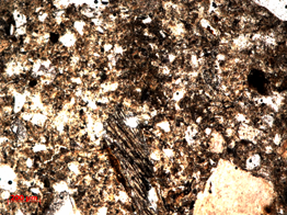
Stone
Characterization of stone samples, through the identification of the minerals they consist of and their crystal* size. According to their optical behaviour under different light source types, the nature of the minerals forming the stone specimen can be determined consulting mineralogical atlases. An optical microscope can also help identify deteriorative agents and weathering products either directly or by micro-chemical testing.
Fluorescence Microscopy is also a useful tool for the identification of areas with microorganism growth and the evaluation of biocide treatments on stone materials.
Wood
Thin sections of wooden samples can be observed under an optical microscope in order to identify the wooden species through their structural characteristics and to compare different species for provenance studies or for building materials comparison.
Optical microscopy (OM) is a technique that allows the magnification of objects through a system of lenses, where naked-eye observation is not sufficient. It is very commonly used in the field of Cultural Heritage, providing morphological and qualitative* information of artefacts during preliminary analysis, throughout conservation operations and after restoration interventions. Produces magnified images of the sample. Depending on the light source and the way it interacts with the specimen, Optical Microscopy can be divided in 4 categories:
Moreover, micro-chemical testing on metallic samples, pigments, metal corrosion patinas or weathering products of stone materials usually requires microscopic observation:
accuracy
time
cost
in situ
invasive
destructive

OM consists in the investigation of a thin- or a cross-section* of the specimen of interest, placed on the microscope’s stage with light aimed at it passing through an opening on the stage or generated from the microscope’s objectives. Light is either transmitted through or reflected by the sample, respectively.