- scanning electron microscopy -
Allows the user to magnify samples through a system of lenses for documentation and preliminary characterization of materials.
dating
morphology
technology
origin
composition
alteration

Ceramic

Glass

Metal

Mortar

Pigments

Stone

Wood
Ceramic Building Materials (CBM)
SEM-EDX can be used for identifying the “chemical signature” of CBM. Tracing elements* both in the clay paste and the aggregates can be an indicator of the geographical origin of ceramic objects by comparison. Surface observation with SEM Imaging can be applied on glazed ceramics to identify the possible degradation areas and to observe the glaze layer diffusion on them.
Glass
Nature, origin, alteration phenomena and degradation state of stained and archaeological glass are usually studied with SEM and SEM-EDX. Alterations, degradation stratigraphy and mapping of elements* for nature and origin studies of glass samples can be performed. Concentrations of some important trace elements* (Titanium, Manganese, iron) for glass provenance studies, are often in the range 0.1–1.0 wt%, which are well detectable with SEM-EDX. Also applied for the identification of the pigments used for glass decoration.
Metals & Alloys
With SEM Imaging it is possible to locate areas of different elemental* composition (chemical mapping of the surface), to observe welding lines that allow the discrimination of different iron-making processes used to obtain the metal and to visualize the chemical phase contrast and features of the metallic grain structure. Stratigraphy of alteration products can also be observed. SEM-EDX is a very useful tool for the identification of alloys, the composition of their main components and impurities. In some cases, the nature of corrosion and weathering products of metals and alloys (patina*) can also be revealed.
Mortars
In mortar characterization SEM-EDX is widely used to identify the type of mortar by chemically analyzing the binder and the aggregates it contains, as well as its hydraulicity index*. It can also be used for comparative studies, comparing the composition of the binder between different mortars in order to understand the provenance of the raw material.
Pigments
By knowing the elemental* composition of a pigment and with supplementary bibliographic research (i.e. Handbooks of Inorganic Pigments) inorganic pigments can be easily identified.
Stone
SEM and SEM-EDX are often used for the morphological and chemical characterization of stone materials. SEM Imaging gives information about the topography of the sample’s surface, allowing the identification of stone alteration products (e.g. black crusts, previous treatment traces, microbiological colonization, cracking, pitting, efflorescence*). With SEM-EDX it is also possible to identify the minerals of which the specimen consists (Bruni et al., 2012). Lastlt SEM-EDX is used for provenance studies: by comparing the elemental* composition of the unknown sample to that of a known one, focusing on the trace elements* which are characteristic of each geographical region, it is possible to have a preliminary indication of the provenance of the stone (quarry).
Wood
To identify wooden species SEM Imaging can also be used in a specific timber characterization procedure, called Wood Anatomy, which consists in the high resolution microscopic observation of the wooden surface’s characteristics (pores, size and density of pores, vessels and fibres) from the three plans of the wood (tangential, radial and longitudinal), which are subsequently compared with keys, tables and atlases.
Scanning Electron Microscopy (SEM) is a technique used for obtaining high-resolution magnified images of surfaces, where Optical Microscopy is not sufficient. When coupled with an Energy Dispersive X-ray (EDS or EDX) spectrometer, the technique allows simultaneous elemental analysis and chemical characterization of a sample’s surface.
Secondary Electron detection (SEM Imaging) Produces a readily interpretable, high-resolution image of the morphology of a sample’s top surface (only a few nm of depth) useful for the observation of different surface characteristics: corrosion product detection, microorganisms, wood anatomy, alterations on the surface, cracks, etc.
Back-scattered Electron detection (SEM-BSE) Produces a readily interpretable, low-resolution image of the morphology of a sample’s top surface the distribution of the different elements in it, useful for semi-quantitative* analysis and chemical mapping if combined with the SEM-EDX method.
accuracy
time
cost
in situ
invasive
destructive
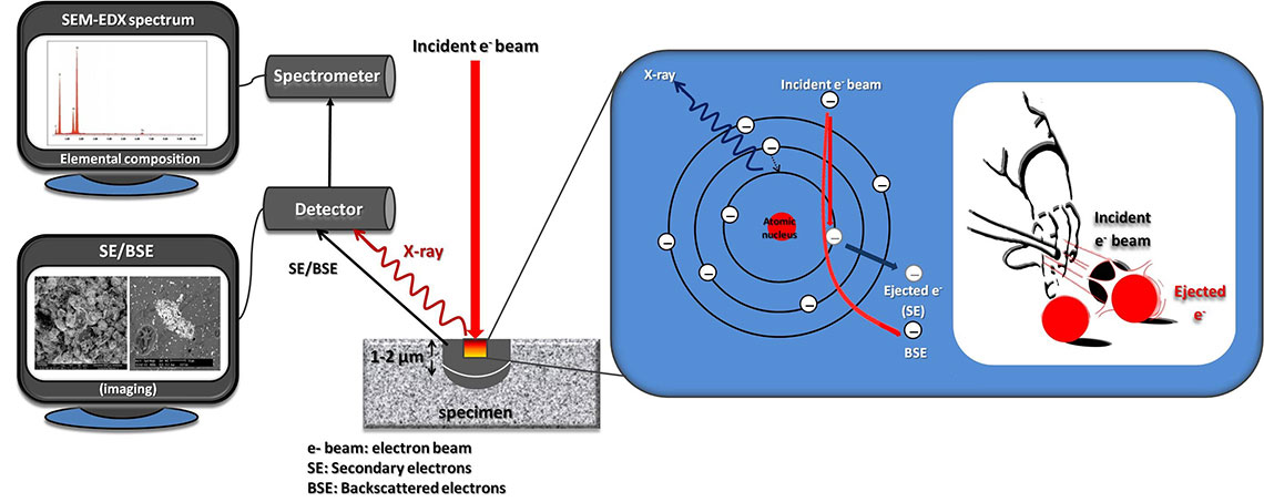
SEM is a type of electron microscopy used to obtain information about the morphology and elemental composition of a sample’s surface. The method consists of bombarding the sample with a high-energy electron beam (Primary radiation). SEM detectors measure the Secondary electrons (SE) and the Backscattered electrons (BSE) emitted, which are characteristic of the element they were emitted from. The detected signal is amplified and converted into digital images.